
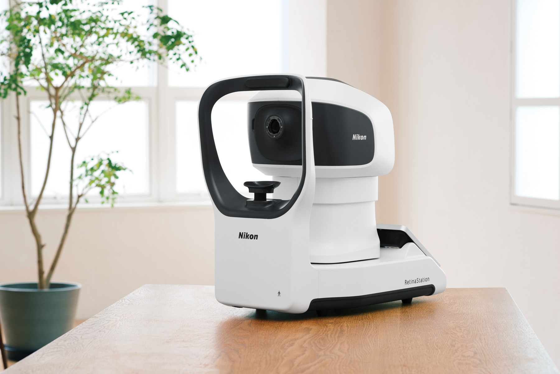
 by Nikon
A Non-Mydriatic Automatic Retinal Camera
- User-Friendly
- Comprehensive Imaging
- Technologically-Advanced
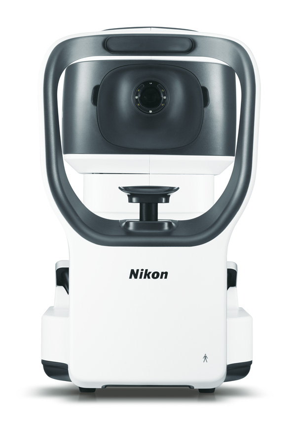
RetinaStation is a standalone, compact, fully-automatic, non-mydriatic fundus camera with image capture and review capabilities. Patient comfort is taken into consideration with the ergonomic chinrest, quick image capture times, and adjustable light intensity to dim the brightness of the flash during capture.
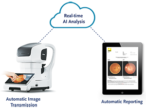
RetinaStation was designed with ease of use, quality imaging, and versatility in mind. With features like 3D tracking alignment, automatic focus, and capture, the RetinaStation, all in one fundus camera will save time during patient workup. Manual acquisition and eye steering for montaged retinal images are also available. RetinaStation also offers access to AI-based Automated Retinal Disease Assessment software* for diabetic retinopathy (DR) and diabetic macular oedema (DMO). Images are analyzed in real-time externally and a comprehensive report is returned. The report is a valuable tool for diabetic retinopathy disease screening, management, and documentation.

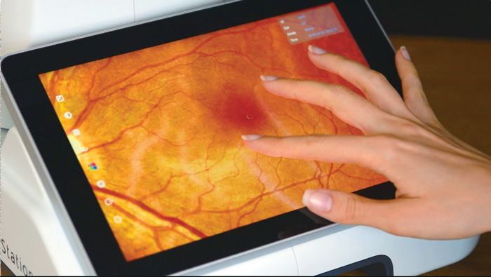
User-Friendly
With a comprehensive, intuitive user interface, a single-tap on the tablet allows the user to automatically align and focus, capture and review fundus images, as well as perform other diagnostic functions. For a closer look at the images, you may simply pinch in or out on the tablet to see a detailed or high-level view. High-resolution image quality is maintained in each view.

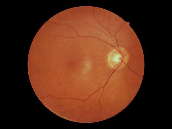
Comprehensive Imaging
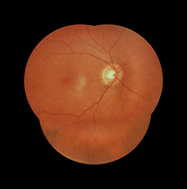
RetinaStation delivers high-resolution color images that are similar to what you would see with direct observation. In addition to retinal images, RetinaStation offers a cornea capture mode for more comprehensive ocular examinations. RetinaStation images can be viewed with Red/Green/Blue channel separation which can be displayed in color or grayscale. Patient data is easily exported or deleted either individually or in multiples.

Technologically-Advanced
The self-contained RetinaStation system enables imaging and editing from a single device. The on-board computer is based on Windows 10 and has several applications that are pre-installed; such as a viewer tool and editing application. It is DICOM compatible therefore interfaces with EMR and PACS systems.
RetinaStation also offers access to AI-based Automated Retinal Disease Assessment software* for diabetic retinopathy (DR) and diabetic macular oedema (DMO). Images are analyzed in real-time externally and a comprehensive report is returned. The report is a valuable tool for diabetic retinopathy disease screening, management, and documentation.
* Contact us for information about software availability in your market.